Difference between macro and micro lens / photography?

Macro and Micro terminology from different companies often creates confusion.
One of our followers on Facebook recently asked us “What is the difference between macro and micro lens / photography?” This was a very good question. When it comes to photography, there is a lot of confusion between the terms macro and micro. The confusion is likely to be attributed to the terminology that is used by different companies to describe their lenses.
1. Difference between Macro and Micro Lens
Macro lenses are lenses that provide at least full life-size magnification (1x magnification). This basically means that if you take a photo of a bee that is 1 inch (2.54cm) in length, the bee will appear as the same size on the camera’s sensor. These lenses are known as ‘Macro’ lenses by Canon. On the other hand, Nikon describes these lenses as ‘Micro’ lenses. So when talking about these types of lenses, the ‘Micro’ and ‘Macro’ terms can be used interchangeably.
2. Difference between Macro and Micro Photography
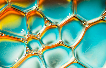
Where you will start seeing some differences is when people talk about ‘macro photography’ and ‘micro photography’. As mentioned above, macro photography is where the subject of our photography appears with a magnification of 1x or more on the camera’s sensor (some people call this 1:1 magnification). Micro photography or photomicrography is relates to photography through a microscope. This is achieved by connecting your camera to the microscope thereby allowing you to capture photos that are 20x the magnification.
If you are interested to see what the photos look like, Nikon currently runs an annual photomicrography competition to recognize the efforts of those involved with photography through the microscope. Here is some information from Nikon International Small World Competition regarding photomicrography –
Photomicrography: The blood-brain barrier in a live zebrafish embryo (20x) – Dr Peters & Dr Taylor from St Jude Children’s Research Hospital.
A photomicrograph is a technical document that can be of great significance to science or industry. But a good photomicrograph is also an image whose structure, color, composition, and content is an object of beauty, open to several levels of comprehension and appreciation.
The subject matter is unrestricted and any type of light microscopy technique is acceptable, including phase contrast, polarized light, fluorescence, interference contrast, darkfield, confocal, deconvolution, and mixed techniques. Entries submitted to Nikon are then judged by an independent panel of experts who are recognized authorities in the area of photomicrography and photography. These entries are judged on the basis of originality, informational content, technical proficiency and visual impact.

Hopefully this has helped you to understand the difference between macro and micro lens and ‘macro photography’ and ‘micro photography’. Here is another entry from the 2012 Nikon International Small World Competition. If you found these photos ‘out of this world’, remember to press LIKE…

Photomicrography: Human bone cancer (osteosarcoma) showing actin filaments (purple), mitochondria (yellow), and DNA (blue) (63x) by Dr Burnette from National Institute of Health.
Photography gear that you may be interested in –